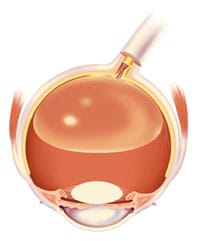
As we age, however, the vitreous thins and separates from the retina. Although this usually results in nothing more than a few harmless floaters, tension from the detached vitreous can sometimes tear the retina.
If liquid were to penetrate the torn area and collect under the retina, the retinal tear can potentially progress to a retinal detachment. A retinal detachment can cause severe, permanent vision loss and requires immediate surgical treatment *by a retinal specialist. Our physician, Dr. Ilyas, has extensive experience in treating retinal tears and detachments.

Indications of retinal tear or detachment may include flashes of light, a group or web of floaters, wavy or watery vision, a sense that there is a veil or curtain obstructing vision, or a sudden drop in the patient’s quality of vision. If you experience any of these symptoms, call us immediately. Early treatment is essential to prevent potential loss of vision. Our retina surgeon can discuss retina surgery options for repair of a retinal tear or detachment and help you find a treatment that best addresses your individual disease.
Macular Hole
A macular hole is exactly what it sounds like: a hole in the macula, the center of the retina responsible for central and reading vision. Specifically, the hole or defect occurs in the fovea, the center of the macula and the most delicate part of the entire retina.
Macular holes almost always develop during the natural aging process, when the vitreous (the gel that fills most of the eye) thins and separates from the macula. This can pull on the macula and cause a hole to form. Less commonly, macular holes are caused by eye injury, intraocular inflammation, retinal detachment and other diseases. Most cases occur in people over the age of 60.

At first, a macular hole may only cause a small blurry or distorted area in the center of vision. As the hole grows over several weeks or months, central vision progressively worsens. Peripheral vision is not affected.
Surgery is over 95% effective for the treatment of macular holes. The procedure is done as an outpatient (no hospital stay required) and performed under local anesthesia. A vitrectomy is performed to remove the vitreous gel. Then, any scar tissue on the macular surface is peeled and removed. Finally, a gas bubble is injected into the eye to help the hole close. As the eye heals, the gas bubble will naturally be removed and replaced by fluid. There is no treatment outside of surgery for macular holes.
To learn more about retina and vitreous diseases and cutting-edge treatments available to you, please call 540-662-1810 today to schedule a consultation.


